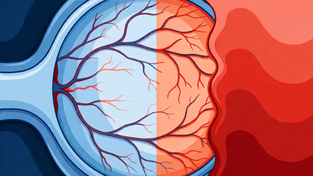
You might think of high blood pressure as a silent killer that only targets your heart or kidneys. But there is another organ quietly suffering under the strain: your eyes. Hypertensive Retinopathy is the medical term for the damage done to the retina’s blood vessels when systemic blood pressure stays too high for too long. It’s not just a minor side effect; it’s a warning sign that your vascular system is struggling, and if ignored, it can lead to permanent vision loss.
Here is the uncomfortable truth: you often won’t notice anything is wrong until significant damage has occurred. According to clinical data, roughly 68% of patients with early-stage hypertensive retinopathy report zero visual symptoms. You could be walking around with narrowed arteries in your eyes, completely unaware, while your risk for stroke and heart attack climbs. This article breaks down exactly what happens inside your eye, how doctors grade the severity, and what you need to do to protect your sight.
The Silent Damage: What Happens Inside the Eye
Your retina is a thin layer of tissue at the back of your eye, packed with light-sensitive cells and tiny blood vessels. These vessels are incredibly delicate. When your blood pressure rises chronically, the walls of these small arteries thicken and narrow to handle the increased force. This process is called arteriolar narrowing. Over time, this reduces blood flow to the retina, starving the tissue of oxygen and nutrients.
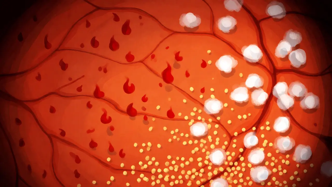
If the pressure remains uncontrolled, the vessel walls become so compromised that they leak. Fluid, lipids (fats), and blood escape into the surrounding retinal tissue. This leakage creates visible signs that an ophthalmologist can spot during a routine exam. You might see flame-shaped hemorrhages, which look like small splashes of ink, or hard exudates, which are yellowish deposits of lipid residue. In more severe cases, you get cotton wool spots-fluffy white patches caused by blocked blood flow leading to localized nerve fiber death.
| Sign | What It Looks Like | Clinical Meaning |
|---|---|---|
| Arteriolar Narrowing | Vessels appear thinner than normal | Early response to high pressure; vessels constrict to protect downstream capillaries. |
| AV Nicking | Veins appear pinched where arteries cross them | Indicates chronic hypertension; stiff arterial walls compress softer veins. |
| Hemorrhages | Red spots or streaks on the retina | Vessel rupture due to excessive pressure; indicates moderate-to-severe disease. |
| Cotton Wool Spots | Fluffy white patches | Micro-infarcts (small areas of tissue death) due to lack of blood supply. |
| Papilledema | Swelling of the optic disc | Sign of malignant hypertension; requires immediate emergency care. |
Grading the Severity: The Keith-Wagener-Barker System
Doctors don’t just say “your eyes look bad.” They use a standardized grading system called the Keith-Wagener-Barker (KWB) classification. This helps determine how urgent treatment needs to be. Understanding your grade gives you a clear picture of your risk level.
- Grade 1: Mild narrowing of the arteries. No bleeding or leakage yet. This is common in people with untreated hypertension for 3-5 years. Many people have no symptoms here.
- Grade 2: More pronounced narrowing and “AV nicking,” where arteries press against veins. This suggests longer-standing hypertension. Vision is usually still normal, but the structural changes are evident.
- Grade 3: Hemorrhages, cotton wool spots, and hard exudates appear. At this stage, you might start noticing blurred vision or blind spots. This indicates significant damage to the retinal barrier.
- Grade 4: All previous signs plus swelling of the optic disc (papilledema). This is a medical emergency often associated with malignant hypertension (BP >180/120 mmHg). Without rapid intervention, permanent blindness is a real threat.
It’s worth noting that even “mild” hypertension (systolic 130-139 mmHg) can cause Grade 1 changes if left untreated for over a decade. The old idea that only severe spikes matter is outdated. Chronic, low-grade elevation does steady damage.
Why Your Eyes Are a Window to Your Heart
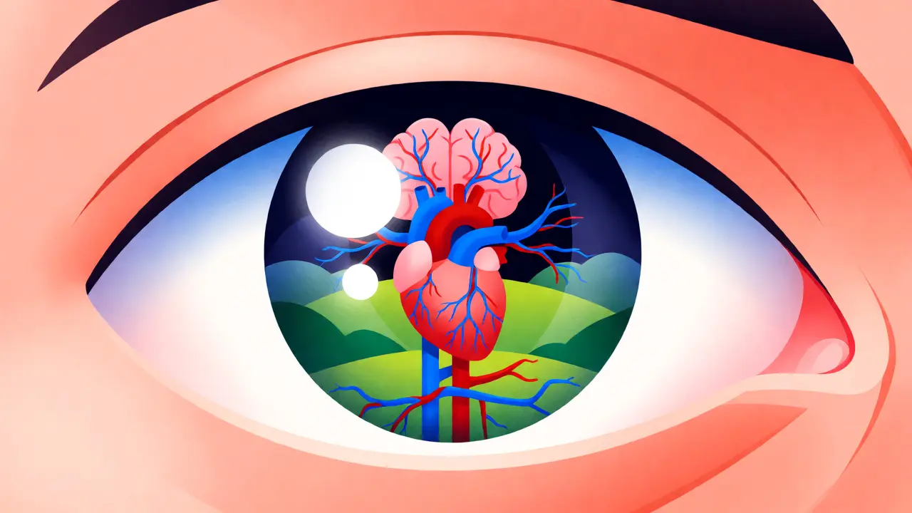
Why should a cardiologist care about your retina? Because the retinal vessels are the only place in your body where doctors can directly observe living blood vessels without surgery. If your retinal vessels are damaged, it’s highly likely that similar damage is happening in your brain, heart, and kidneys.
Studies show a direct correlation between retinopathy severity and systemic health risks. For instance, patients with Grade 4 retinopathy have a 78% higher risk of stroke compared to those without these eye changes. Furthermore, specific findings like AV nicking are linked to a 3.2x higher risk of stroke. This makes hypertensive retinopathy a powerful predictor of overall cardiovascular mortality. If your eye doctor flags these signs, it’s not just about saving your sight-it’s about preventing a heart attack or stroke.
Consider this scenario: A patient comes in complaining of occasional headaches and blurry vision. Their blood pressure reads 165/105 mmHg. An eye exam reveals Grade 3 retinopathy. This finding confirms that their hypertension has been uncontrolled for years, not just recently. It shifts the treatment plan from simple lifestyle advice to aggressive medication management and closer monitoring of kidney function.
Symptoms You Should Not Ignore
While early stages are asymptomatic, advanced hypertensive retinopathy produces distinct symptoms. Don’t wait for total blindness to seek help. Watch out for:
- Blurred Vision: Often the first noticeable sign, especially if it affects central vision due to macular edema (fluid buildup in the center of the retina).
- Dark Spots or Floaters: Caused by hemorrhages leaking into the vitreous humor (the gel-like substance filling the eye).
- Sudden Vision Loss: In cases of malignant hypertension, you might experience a “curtain” falling over part of your visual field. This can happen within hours of a BP spike.
- Double Vision: Less common, but can occur if high pressure affects the nerves controlling eye movement.
A common mistake patients make is assuming these symptoms are just aging or dry eyes. However, sudden changes, especially combined with high blood pressure readings, warrant an immediate call to your healthcare provider. Data suggests that 42% of patients delay seeking treatment until vision deterioration impacts daily activities, leading to an average diagnosis delay of 18-24 months after symptoms begin.
Diagnosis and Modern Technology
Gone are the days when diagnosis relied solely on a doctor peering through a magnifying lens. Today, technology plays a huge role. Optical Coherence Tomography (OCT) is now standard in many clinics. It uses light waves to take cross-section pictures of your retina, detecting thickness changes and fluid accumulation invisible to the naked eye. OCT can show retinal thickness increases of 10-15% during exudative phases, providing objective data to track progress.
Artificial Intelligence is also entering the space. Systems like IDx-DR and newer AI tools can screen for retinopathy with over 90% accuracy. This is crucial because primary care physicians are increasingly integrating retinal screening into routine checkups. If you haven’t had a dilated eye exam in over a year, ask your GP if a referral to an ophthalmologist is warranted, especially if you have Stage 2 hypertension.
Treatment: Controlling the Pressure to Save the Sight
The good news? Hypertensive retinopathy is largely reversible if caught before permanent scarring occurs. The primary treatment isn’t eye drops or surgery-it’s lowering your blood pressure.
For acute cases, such as malignant hypertension, reducing systolic BP by 25 mmHg within 24-48 hours can resolve retinal changes in 65% of cases. For chronic management, consistent control is key. Most patients see vision improvement within 7-10 days of stabilizing their BP. However, if the macula (the center of vision) was damaged, recovery can take 3-6 months, and some visual field defects may remain permanent.
Medication choice matters too. Recent guidelines suggest that ACE inhibitors may reduce retinal damage progression by 32% compared to calcium channel blockers. Always discuss your specific medication regimen with your doctor, considering other conditions like diabetes, which multiplies the risk of vision loss by nearly five times.
Prevention and Long-Term Management
Prevention beats cure every time. Here is your action plan:
- Monitor at Home: Invest in a validated home blood pressure monitor. Learn to use it correctly (rested, feet flat, arm supported). Consistency is more important than single readings.
- Annual Dilated Exams: Even if your vision feels fine, get your eyes checked annually if you have hypertension. Early detection of Grade 1 changes allows for timely intervention.
- Lifestyle Changes: Reduce sodium intake, maintain a healthy weight, and engage in regular aerobic exercise. These steps lower BP naturally and improve vascular health.
- Adhere to Medication: Skipping doses causes fluctuations that stress vessel walls. Studies show 70% adherence rates when patients understand the link between meds and eye health.
Remember, your eyes are telling you a story about your entire circulatory system. Listening to that story can save your sight and potentially your life.
Can hypertensive retinopathy be cured?
Yes, in early stages. If blood pressure is controlled promptly, retinal changes like hemorrhages and exudates can resolve, and vision often returns to normal. However, severe damage involving the optic nerve or macular scarring may result in permanent vision loss.
How quickly does high blood pressure affect the eyes?
Retinal damage can begin after just 3-5 years of uncontrolled hypertension. However, acute changes like papilledema can occur within 48-72 hours during a hypertensive crisis (BP >180/120 mmHg).
Is hypertensive retinopathy the same as diabetic retinopathy?
No, though they share similarities like hemorrhages and exudates. Diabetic retinopathy is caused by high blood sugar damaging vessels, while hypertensive retinopathy is caused by high blood pressure. Patients with both conditions face a significantly higher risk of vision loss.
Do I need special glasses for hypertensive retinopathy?
Glasses correct refractive errors but do not treat retinopathy. Treatment focuses on lowering blood pressure. In rare cases of macular edema, anti-VEGF injections or laser therapy might be used, but BP control is the primary remedy.
How often should I get my eyes checked if I have high blood pressure?
For stable hypertension, annual exams are recommended. If you have resistant hypertension or existing retinopathy, your doctor may recommend screenings every 6 months to monitor for progression.
Neil Martin
September 5, 2026 AT 14:02It is frankly insulting that most primary care physicians treat hypertension as a mere number to be managed rather than a systemic vascular catastrophe. The arteriolar narrowing described here is not just a local phenomenon; it is the canary in the coal mine for impending cerebrovascular accidents. If you are not getting a dilated fundoscopic exam annually, you are essentially flying blind regarding your true cardiovascular risk profile. Most patients ignore this until they have Grade 3 changes, at which point the micro-infarcts are already cemented into their visual field. Stop relying on generic advice and demand specific retinal imaging if your systolic pressure has hovered above 140 for more than five years.
Kim Pender
September 6, 2026 AT 08:57I had no idea my blurry vision was linked to my blood pressure meds not working right.
My doctor never mentioned the eye thing.
Neil Sahli
September 8, 2026 AT 08:13Oh wow, another reason why ignoring your health is basically volunteering for early retirement from life! 🎉
The fact that 68% of people have zero symptoms is absolutely hilarious because we all think we're invincible until our eyes start leaking fluid. But hey, at least the treatment is simple: take your pills and don't eat salt like it's candy. You can totally fix this before you end up needing a guide dog. Just do the work, seriously, it's not rocket science, it's just basic self-preservation!
Pramay Dattani
September 9, 2026 AT 11:16This is typical western over-diagnosis. In India we know that stress and diet cause these issues, not just some magic pill. My uncle had high BP and his eyes were fine because he ate turmeric and walked daily. American doctors always want to sell you expensive tests like OCT when simple observation works. Why do US citizens need so many machines? It is waste of money. We should focus on natural remedies instead of grading severity with fancy names like Keith-Wagener-Barker. Typical arrogant medical establishment.
Aurelio Haney
September 12, 2026 AT 09:14Grading the severity is crucial but people get too hung up on the labels. 😒
Grade 1 is common, sure, but if you aren't fixing the root cause, you're just waiting for Grade 4. Don't wait for papilledema to panic. Fix your lifestyle now or suffer later. :/
Falgun R Patel
September 13, 2026 AT 06:26There is a profound philosophical implication in the statement that the retina is the only place we can observe living blood vessels directly. It suggests that our perception of reality is literally tied to the health of our vascular system. If the lens through which we view the world is clouded by pressure, how much of our experience is distorted?
We must approach this not just as a medical issue but as an existential one. Protecting your sight is protecting your ability to engage with the world meaningfully. Be gentle with yourself during treatment, but be rigorous with prevention. The journey to better health is a marathon, not a sprint, and every small step counts towards clarity.
Mary Tait
September 14, 2026 AT 08:26This article is clearly biased toward Western medical practices. In America, we prioritize aggressive intervention, which is correct. The correlation between Grade 4 retinopathy and stroke risk is undeniable evidence of superior diagnostic capability. Patients who delay seeking treatment due to ignorance deserve the consequences. Our healthcare system, while imperfect, provides the best outcomes for those who follow protocol strictly. Do not let alternative medicine enthusiasts confuse you with anecdotes; data wins.
Rebecca Makayla Crane
September 14, 2026 AT 14:51OCT scans are so last season, darling. 🙄 We’re moving toward AI-driven diagnostics that can predict retinal thickness changes before they even happen. 💅
If you’re still doing manual fundoscopy, you’re basically practicing archaeology. Get the tech or get left behind. 📉✨