Imaging Test Decision Tool for Urinary Retention
Select Your Clinical Scenario
This tool helps determine the most appropriate imaging test based on your clinical presentation and concerns.
When the bladder won’t empty properly, the discomfort can range from mild urgency to painful swelling. Figuring out why this happens often means looking beyond symptoms and straight into the body with imaging. Below, we break down which scans work best, how they’re used, and what pitfalls to watch out for.
Key Takeaways
- Ultrasound is the first‑line test for most urinary‑retention cases because it’s quick, safe, and cheap.
- CT scans excel at spotting stones, tumors, or severe blockages that ultrasound might miss.
- MRI provides the most detailed view of soft‑tissue problems such as prostate enlargement or neurogenic causes.
- Choosing the right test depends on patient history, suspected cause, and available resources.
- Sometimes imaging must be paired with urodynamic studies or lab tests for a full picture.
Urinary retention is a condition where the bladder cannot completely empty, leading to a buildup of urine. It can be acute (sudden onset, often painful) or chronic (gradual, sometimes unnoticed). Common triggers include prostate enlargement, urethral strictures, bladder stones, neuro‑genic disorders, and certain medications.
Why Imaging Matters
Symptoms alone rarely tell you what’s blocking the flow. Physical exams and basic labs might hint at a problem, but they can’t show the exact location or size of a blockage. Imaging fills that gap by providing a visual map of the urinary tract, helping clinicians decide whether surgery, medication, or watchful waiting is appropriate.
Common Imaging Modalities
Each scan type has strengths and limits. Below we introduce the four most frequently used tests.
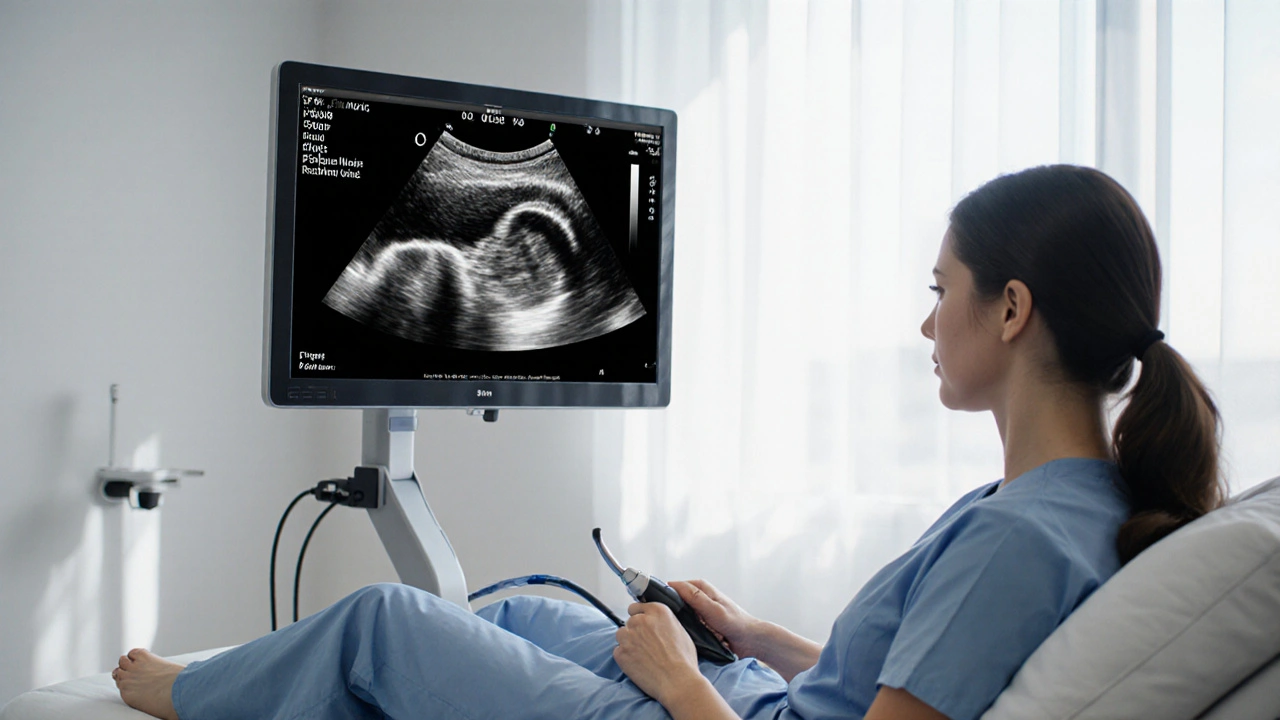
Ultrasound
Ultrasound is a real‑time, non‑invasive technique that uses high‑frequency sound waves to create images of the kidneys, ureters, and bladder. It can measure post‑void residual volume, detect hydronephrosis, and locate large stones. Because it involves no radiation, it’s safe for repeat use and for pregnant patients.
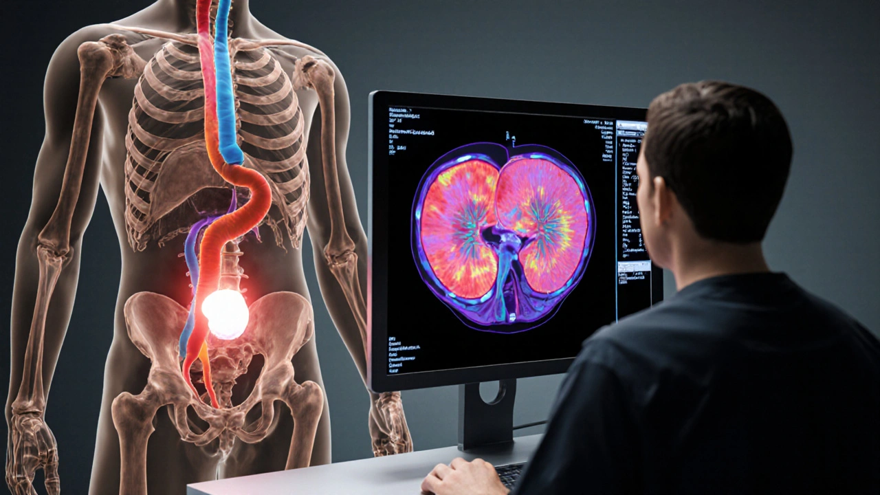
CT Scan
CT (computed tomography) scan combines multiple X‑ray images to generate cross‑sectional views. Modern low‑dose protocols keep radiation under 5mSv for a typical abdomen‑pelvis study. CT is excellent for spotting small calculi, complex tumors, or severe anatomical anomalies that may be invisible on ultrasound.
MRI
MRI (magnetic resonance imaging) uses magnetic fields and radio waves to produce high‑resolution images of soft tissue. It’s the gold standard when clinicians suspect neuro‑genic causes, prostate cancer invasion, or inflammatory conditions. MRI avoids ionising radiation but costs more and takes longer.
X‑ray (KUB)
KUB (Kidney‑Ureter‑Bladder) X‑ray provides a quick, low‑cost snapshot of the abdomen. While it can reveal large stones and certain calcifications, it’s rarely the sole modality for urinary retention because it lacks soft‑tissue detail.
Choosing the Right Test: Decision Factors
| Modality | Best For | Strengths | Limitations | Typical Cost (UK) |
|---|---|---|---|---|
| Ultrasound | Post‑void residual volume, hydronephrosis, large stones | Fast, no radiation, bedside availability | Operator‑dependent, limited for deep pelvic structures | £80‑£120 |
| CT Scan | Small stones, complex tumors, detailed anatomy | High spatial resolution, quick acquisition | Ionising radiation, may need contrast | £250‑£350 |
| MRI | Soft‑tissue lesions, neuro‑genic causes, prostate assessment | Excellent soft‑tissue contrast, no radiation | Longer scan time, higher cost, contraindicated with certain implants | £500‑£700 |
| KUB X‑ray | Large calculi, quick screening | Very low cost, widely available | Poor soft‑tissue detail, limited sensitivity | £30‑£50 |
In practice, doctors start with ultrasound because it’s safe and cheap. If ultrasound can’t explain the retention-say the residual volume is high but no obstruction appears-then a CT or MRI is ordered based on the suspected cause.
Step‑by‑Step Diagnostic Pathway
- Take a detailed history (symptom onset, pain, medication, recent surgeries).
- Perform a physical exam, focusing on the abdomen and prostate (for men).
- Measure post‑void residual volume with a bedside bladder scan.
- If residual >200mL or symptoms are severe, order a urinary retention imaging ultrasound.
- Review ultrasound:
- Hydronephrosis present? → Consider obstructive cause.
- Large bladder wall thickening? → Think infection or neuro‑genic issue.
- If ultrasound is inconclusive, select a second‑line test:
- CT for suspected stones or tumours.
- MRI for prostate cancer, spinal cord lesions, or complex soft‑tissue disease.
- Combine imaging findings with lab results (urinalysis, PSA, electrolytes) to pinpoint the cause.
- Plan treatment: catheterisation, medication, surgical intervention, or referral to a urologist.

Pitfalls & Practical Tips
- Operator skill matters. A novice sonographer may miss a small ureteric stone; ask for a repeat scan if symptoms persist.
- Contrast allergies can limit CT usefulness. Non‑contrast CT is still very effective for stones.
- Patients with claustrophobia may struggle with MRI; mild sedation or an open‑MRI scanner can help.
- Always correlate imaging with clinical signs. A stone seen on CT may be incidental if the bladder still empties adequately.
- Consider radiation dose, especially for younger patients. Opt for ultrasound or MRI when feasible.
When Imaging Isn’t Enough
Sometimes the picture remains hazy even after multiple scans. In those cases, urodynamic studies-tests that measure pressure and flow during urination-provide functional data that imaging can’t. Blood tests for calcium, phosphate, and renal function help rule out metabolic stone formation. Finally, cystoscopy (a direct look inside the bladder) can detect internal strictures or tumors missed by external imaging.
Frequently Asked Questions
Can a simple ultrasound miss the cause of urinary retention?
Yes. Ultrasound is great for spotting large stones and bladder overflow, but tiny ureteric stones, deep pelvic masses, or neuro‑genic issues may be invisible. That’s why a CT or MRI is often ordered if the ultrasound doesn’t match the clinical picture.
Is radiation from CT scans a major concern?
A typical abdomen‑pelvis CT delivers about 5-10mSv, roughly the amount of natural background radiation a person receives in 1‑2years. For most adults, the diagnostic benefit outweighs the risk, but doctors try to limit repeat scans and use low‑dose protocols, especially in younger patients.
When should I expect a referral to a urologist?
If imaging shows a clear obstruction (large stone, tumour, severe prostate enlargement) or if symptoms persist despite catheter drainage, a urologist should be involved promptly. Chronic retention that leads to kidney damage also warrants specialist care.
Can MRI be used without contrast for urinary problems?
Yes. Non‑contrast MRI can still delineate soft‑tissue anatomy, detect prostate enlargement, and assess spinal cord lesions that may cause neuro‑genic retention. Contrast is added when vascular details or tumour perfusion need clarification.
Neviah Abrahams
October 12, 2025 AT 12:39Seeing the decision tool parade itself as a miracle cure for urinary retention feels like a bad joke. The flow of options jumps around like a nervous cat, leaving clinicians scrambling for a clear path. It promises lightning‑fast answers but drops crucial nuance about patient history. In the end the tool might do more harm than good by oversimplifying complex cases.
Uju Okonkwo
October 12, 2025 AT 14:02Hey, I get where you’re coming from – the tool can seem hectic at first. Think of it as a flexible guide that you can tweak to each patient’s story. Start with the simple ultrasound tip, then let the algorithm point you toward CT or MRI only if the first scan isn’t enough. It’s okay to pause, re‑evaluate, and involve a urologist when things stay blurry.
Destiny Hixon
October 12, 2025 AT 15:25Look folks the US healthcare loves flashy tech but forgets good old common sense. This “high‑tech” decision maker just tells you to get an MRI when you could've done a cheap ultrasound. Stop wasting dollars and stick to what works.
mike brown
October 12, 2025 AT 16:49Actually, throwing out advanced imaging isn’t fair – some cases need the detail only CT or MRI can give. Just because it costs more doesn’t mean it’s useless. Balance is key, not blind dismissal.
Brett Coombs
October 12, 2025 AT 18:12They don’t want you to know how much radiation they’re putting in you. The CT scans in that tool are a trap, hidden danger for the public.
John Hoffmann
October 12, 2025 AT 19:35The claim that computed tomography inevitably exposes patients to hidden danger is a mischaracterization of modern imaging protocols.
Contemporary low‑dose CT algorithms reduce the effective dose to levels comparable to several months of natural background radiation.
Moreover, the diagnostic benefit of identifying a obstructing calculus or a neoplastic lesion often outweighs the modest risk associated with a single scan.
It is also important to recognize that ultrasound, while radiation‑free, suffers from operator dependency and limited visualization of deep pelvic structures.
When the clinical picture suggests a small stone or subtle soft‑tissue abnormality, ultrasound may simply miss the pathology.
In such scenarios, a targeted CT provides high‑resolution cross‑sectional images that can change management decisions.
The article’s decision tool correctly prioritizes CT for acute stone detection, reflecting current evidence‑based practice.
Radiation concerns should be addressed by employing dose‑modulation techniques and limiting scan length to the region of interest.
Patients with a history of multiple scans can also benefit from cumulative dose tracking, which many institutions now incorporate into electronic health records.
Furthermore, the notion that “CT is always a trap” ignores the fact that certain malignancies are only apparent on contrast‑enhanced CT.
The tool’s inclusion of alternative imaging, such as MRI for neuro‑genic causes, demonstrates an awareness of radiation‑free options when appropriate.
However, the decision algorithm could be improved by explicitly mentioning low‑dose protocols for stone assessment.
Clinicians should also consider patient‑specific factors such as age, pregnancy status, and renal function when selecting an imaging modality.
Ultimately, the choice between ultrasound, CT, and MRI must be individualized, balancing diagnostic yield against potential hazards.
A nuanced discussion with the patient about the risks and benefits remains essential for informed consent.
Shane matthews
October 12, 2025 AT 20:59In practice the first step is usually a quick bedside bladder scan – it’s fast, painless, and gives you the post‑void residual volume straight away.
Rushikesh Mhetre
October 12, 2025 AT 22:22Exactly! A bedside scan can save you a trip to radiology and cut down on unnecessary radiation exposure!!! If the residual is high, you’ve got a clear sign to dig deeper with ultrasound or CT – whichever fits the patient’s story best!!! Keep the momentum going, and don’t let the fear of “missing something” stall your decision!!!
Sharath Babu Srinivas
October 12, 2025 AT 23:45Ultrasound remains the workhorse for initial evaluation – it’s non‑invasive, cost‑effective, and provides real‑time volume measurements 🚀. However, remember its limitations: deep‑pelvic lesions and tiny calculi can elude detection 🙈.
Halid A.
October 13, 2025 AT 01:09Thank you for highlighting both strengths and caveats. In clinical pathways, an initial ultrasound followed by targeted CT when suspicion persists aligns with evidence‑based guidelines. This stepwise approach optimizes resource utilization while maintaining diagnostic accuracy.
Brandon Burt
October 13, 2025 AT 02:32Honestly, the whole decision tool feels like it was cobbled together by someone who skimmed a textbook and thought a few drop‑downs would replace a thorough clinical assessment. The user interface is clunky, the language is overly generic, and there’s no mention of how to handle patients with multiple comorbidities that could affect imaging choices. It also glosses over the fact that many hospitals don’t have immediate access to open‑MRI suites, which makes the “claustrophobia” adjustment practically irrelevant for a large portion of the population. While the inclusion of cost considerations is a nice touch, the cost ranges are UK‑centric and may mislead readers in other healthcare systems. In short, the tool could benefit from more nuanced scenarios, clearer explanations of each modality’s diagnostic yield, and a greater awareness of real‑world resource constraints.
Gloria Reyes Najera
October 13, 2025 AT 03:55Look the tool oversimplifies but the reality is you need to factor in patient age and insurance coverage before picking MRI over CT. It’s not just a click.
Gauri Omar
October 13, 2025 AT 05:19When you stare at a bladder that won’t empty, the frustration builds like a storm, and you need imaging that cuts through the gloom. The guide tries to be helpful but sometimes it feels like it’s whispering in a thunderstorm.
Willy garcia
October 13, 2025 AT 06:42Don’t let the storm overwhelm you – start with a simple scan, evaluate the numbers, and then decide if you need the heavy‑duty imaging. It’s a steady climb, not a leap.
zaza oglu
October 13, 2025 AT 08:05Hey folks, think of the imaging ladder as a rainbow of options – ultrasound is the bright yellow base, CT shines orange for stones, and MRI glows deep blue for soft‑tissue mysteries!!! Use each color wisely and your patients will thank you!!!
Vaibhav Sai
October 13, 2025 AT 09:29Love the rainbow metaphor! It really paints a clear picture of when to reach for each modality – bright and vivid!!! Just remember the rainbow ends where resources begin, so always check availability before you chase the colors.
Lindy Swanson
October 13, 2025 AT 10:52Well, I guess if you’re happy with guessing, the tool isn’t for you.